
We characterized and validated the protocol using different methods, including Fluorescence-Lifetime Imaging Microscopy (FLIM) and Raman spectroscopy. MAGIC is a methodology that permits, with a glycerol-based procedure, to enhance the autofluorescence of myelin of fixed samples. To overcome these limitations, we developed MAGIC (Myelin Autofluorescence imaging by Glycerol Induced Contrast enhancement), a simple label-free method that opens the possibility of performing sub-micron resolution fluorescence imaging of myelinated fibers in 3D at the mesoscale level of different mammalian brains. Moreover, the necessity of prelabeled conditions prevents the possibility of studying archived samples. Nevertheless, with these approaches the analysis is confined to animal models where gene manipulation can be performed and virus/dyes can be administrated in vivo. Transgenic models 12, injection of virus 13, 14 or injection of dyes 15 can be used for specific labelling of circuits of interest. Exogenous dyes 11 are used to stain fibers composing the white matter, but nonspecific binding and inefficient diffusion of dyes hinder single fiber imaging in large volumes. In this respect, myelin staining is an unmet technical challenge. However, they need a source of contrast to detect the structure of interest.
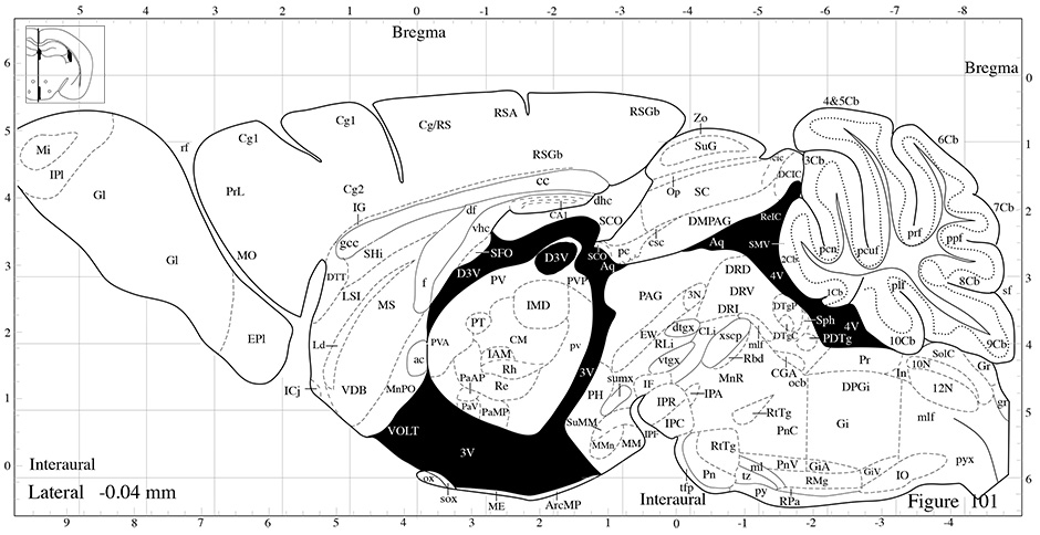
MOUSE HIPPOCAMPUS ANATOMY LABELS SERIAL
For instance, cutting-edge microscopy techniques such as micro-optical sectioning tomography (MOST) 5, 6, serial two-photon tomography (STP) 7, block-face serial microscopy (FAST) 8, and light sheet microscopy (LSM) 9, 10, permit the reconstruction of large volumes of tissue and even entire organs with micrometer resolution. Optical methods have the potential for scalable large-area high-resolution mapping. Several methods have been developed to map the network of interneuron connections still, they are limited by the volume that they can analyze (Electron Microscopy) 2, by the spatial resolution achievable (MRI) 3 or by the sophisticated equipment needed for the measurement (CARS, THG microscopy) 4. Reconstructing the intricate organization of these connections remains a challenging goal to achieve due to technical limitations, especially on human brain tissues. Most long-range projecting axons are wrapped by the myelin sheath to permit a reliable and efficient signal transmission 1. The organization of nerve fibers spans many orders of organization: from the microscale of the single axon to the macroscale of the long-range connections connecting far-distant brain regions. Individual neurons are interconnected to hundreds or even thousands of other cells forming networks that can extend over large volumes, up to several centimeters for the human brain. The brain shows a complex network of neurons, capable of storing and processing information from a myriad of different inputs regulating cognitive processes and behavior. Our study provides a novel method for 3D label-free imaging of nerve fibers in fixed samples at high resolution, below micrometer level, that overcomes the limitation related to the myelinated axons exogenous labeling, improving the possibility of analyzing brain connectivity. We demonstrate its broad applicability by performing mesoscopic reconstruction at a sub-micron resolution of mouse, rat, monkey, and human brain samples and by quantifying the different fiber organization in control and Reeler mouse's hippocampal sections. We exploit the high axial and radial resolution of two-photon fluorescence microscopy (TPFM) optical sectioning to decipher the mixture of various fiber orientations within the sample of interest. Here, we propose MAGIC (Myelin Autofluorescence imaging by Glycerol Induced Contrast enhancement), a tissue preparation method to perform label-free fluorescence imaging of myelinated fibers that is user friendly and easy to handle. The possibility of discriminating between different adjacent myelinated axons gives the opportunity of providing more information about the fiber composition and architecture within a specific area. Different technologies try to address this goal however, they are limited either by the ineffective labeling of the fibers or in the achievable resolution. Analyzing the structure of neuronal fibers with single axon resolution in large volumes is a challenge in connectomics.


 0 kommentar(er)
